


Disease
Advanced Knee Recovery Solutions
Etiology
Meniscal Injury
Meniscal injuries occur in various sports, especially contact sports, and are also relatively common in daily activities and work, often combined with other ligament injuries. The medial meniscus is easily injured when the lower leg rotates externally relative to the femur, while the lateral meniscus is more prone to injury during internal rotation of the tibia. Meniscal injuries can also occur due to hyperflexion or hyperextension of the knee or direct impact between the femur and tibia. Reports from abroad indicate that medial meniscus injuries are five times more prevalent than lateral meniscus injuries, while domestic reports suggest a higher prevalence of lateral meniscus injuries.
Medial Collateral Ligament (MCL) Injury
The MCL consists of superficial and deep layers without a distinct gap between them. The superficial layer originates near the adductor tubercle and inserts on the inner side of the upper end of the tibia, while the deep layer starts at the medial epicondyle and inserts on the inner side of the upper end of the tibia, contributing to the joint capsule and connecting to the medial meniscus. Injuries to the MCL stem from forces acting from the outside, such as tibial abduction and external rotation or femoral adduction and internal rotation.
Lateral Collateral Ligament (LCL) Injury
LCL injuries are less common and typically result from the application of force to the inside of the knee joint or other causes leading to knee joint varus injury, often accompanied by injuries to the joint capsule, peroneal muscles, biceps femoris, hamstring muscles, or even the common peroneal nerve.
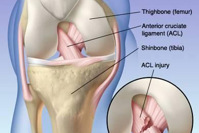
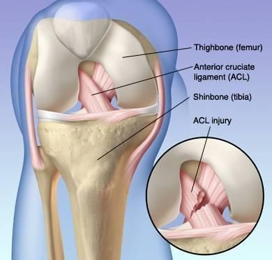
Anterior Cruciate Ligament (ACL) Injury
The ACL starts between the anterior areas of the tibial condyle and the anterior horn of the lateral meniscus, ending at the inner part of the lateral femoral condyle. It consists of the posterior lateral bundle and the anterior medial bundle. ACL injuries are more common, often part of combined injuries, but can also occur as isolated injuries.
Posterior Cruciate Ligament (PCL) Injury
The PCL attaches to the posterior aspect of the tibial joint surface, extends to the posterior upper end of the tibia, runs behind the posterior medial bundle of the anterior cruciate ligament, and ends at the lateral aspect of the medial femoral condyle. The PCL is relatively robust, and thus injuries are less frequent, usually resulting from significant external forces and often accompanied by other injuries.
Examination
Clinical Examination: Includes symptom observation, joint stability assessment, measurement of joint range of motion, etc.
Imaging Studies: X-rays, MRI, CT scans, etc., used to view bone structures, soft tissue, and the extent of injuries.
Arthroscopic Examination: Directly observes the internal conditions of the joint, aiding in diagnosis and treatment.
Diagnosis
Meniscal Injury
Patients often have a history of trauma, immediate post-injury pain, subsequent knee joint swelling, inaccurately localized acute pain, and later, specific site pain. Post-injury, joint effusion occurs, along with joint locking and the appearance of "giving way" during knee joint movement, accompanied by clicking, and palpable localized tenderness in the joint space. McMurray's Test is commonly positive and is the most commonly used examination method. The Apply Test can induce pain on the injured side and assesses the situation during squatting under load. Some also perform a swinging test, placing one thumb in the injured side's joint space and gently swaying the leg, feeling the meniscus moving in and out of the space, which is positive if accompanied by pain.
Knee joint arthrography is a frequently used diagnostic tool, aiding in injury localization. Although still used in some cases, it has gradually been replaced by newer examination methods. Arthroscopic examination has a confirmation rate of up to 90% and can be used for surgery but has limitations in observing the posterior horn of the medial meniscus. MRI is valuable for diagnosing joint soft tissue injuries.
Medial Collateral Ligament (MCL) Injury
Post-injury, intense pain occurs on the medial side of the knee joint, relieved and then exacerbated, leading to medial swelling and ecchymosis. At 30° of knee flexion, an abnormal joint space opening sensation can be felt, reduced MCL tension, and positive valgus stress test. Performing valgus stress X-rays for bilateral comparison shows increased joint space on the affected side by over 10°, indicating a complete rupture of the MCL, along with potential anterior cruciate ligament injury. MRI provides a clearer diagnosis.
Lateral Collateral Ligament (LCL) Injury
Patients often have a history of an inside force on the knee joint, post-injury lateral knee pain, swelling, and significant local tenderness, frequently with fibular head fractures. When accompanied by injuries to adjacent structures, corresponding symptoms occur. Valgus stress is positive, reduced LCL tension, and palpable tenderness and an abnormal opening sensation. Valgus stress X-rays show increased joint space on the affected side.
Anterior Cruciate Ligament (ACL) Injury
ACL injuries often occur due to acute knee joint trauma, perceived tearing sensation, knee joint pain, instability, inability to repeat movements, or continue exercising. Subsequent joint swelling and hemarthrosis occur, and the drawer test is positive. Acute severe pain often makes detailed examinations challenging, but examinations can be conducted after anesthesia or in the post-acute period. Positive anterior drawer test, Lachman test, pivot shift test, and jerk test indicate ACL injury. The Lachman test performed with the patient's leg hanging is positive, indicating ACL injury. X-rays for detection of avulsion fractures are diagnostically meaningful. Simultaneous anterior drawer test X-rays show anterior tibial displacement, indicating ACL injury. MRI provides relatively accurate diagnosis.
Posterior Cruciate Ligament (PCL) Injury
Symptoms of PCL injuries resemble those of ACL injuries and have a clear history of acute trauma. The posterior drawer test is positive, and X-rays can be performed for diagnosis. Patients lying supine with both feet on the examination table at approximately 90° of knee flexion may experience a posterior tibial sag; when the examiner holds the patient's femur distally and flexes the hip and knee, the posterior movement of the proximal tibia is more pronounced, indicating PCL rupture. X-rays show PCL avulsion fractures. MRI is relatively accurate for diagnosing PCL injuries.